T Cell Activation Bioassays
Novel biologics formats, including bispecific antibodies and CAR-T cell therapies, require robust, reproducible measurement of T cell activation. Our ICH-prequalified bioassays enable simple, scalable solutions for drug screening and development.
These assays consist of a genetically engineered T cell line that expresses a luciferase reporter driven by either an NFAT response element or an IL-2 promoter. When engaged with either an anti-TCR/CD3 stimulus alone or an anti-TCR/CD3 and an anti-CD28 stimulus, receptor-mediated signaling induces luminescence.
Filter By
Shop all T Cell Activation Bioassays
Showing 0 of 3 Products
Featured Publications
T Cell Activation Bioassay (IL-2)
Wu, L. et al. (2020) Trispecific Abs enhance the therapeutic efficacy of tumor-directed T cells through T cell receptor co-stimulation. Nat. Cancer 1, 86–98. doi: 10.1038/s43018-019-0004-z
T Cell Activation Bioassay (NFAT)
Sahin, U. et al. (2020) An RNA vaccine drives immunity in checkpoint-inhibitor-treated melanoma. Nature 585, 107–112. doi: 10.1038/s41586-020-2537-9
Nakazawa, H. et al. (2020) Association behavior and control of the quality of cancer therapeutic bispecific diabodies expressed in E. coli. Biochem. Eng. J. 160, 107636. doi: 10.1016/j.bej.2020.107636
An Introduction to T Cell Activation
T cells play a central role in cell-mediated immunity and can regulate long-term, antigen-specific, effector and memory responses. Immunotherapy strategies aimed at inducing, strengthening and/or engineering T cell responses have recently emerged as promising approaches for the treatment of diseases such as cancer and autoimmunity.
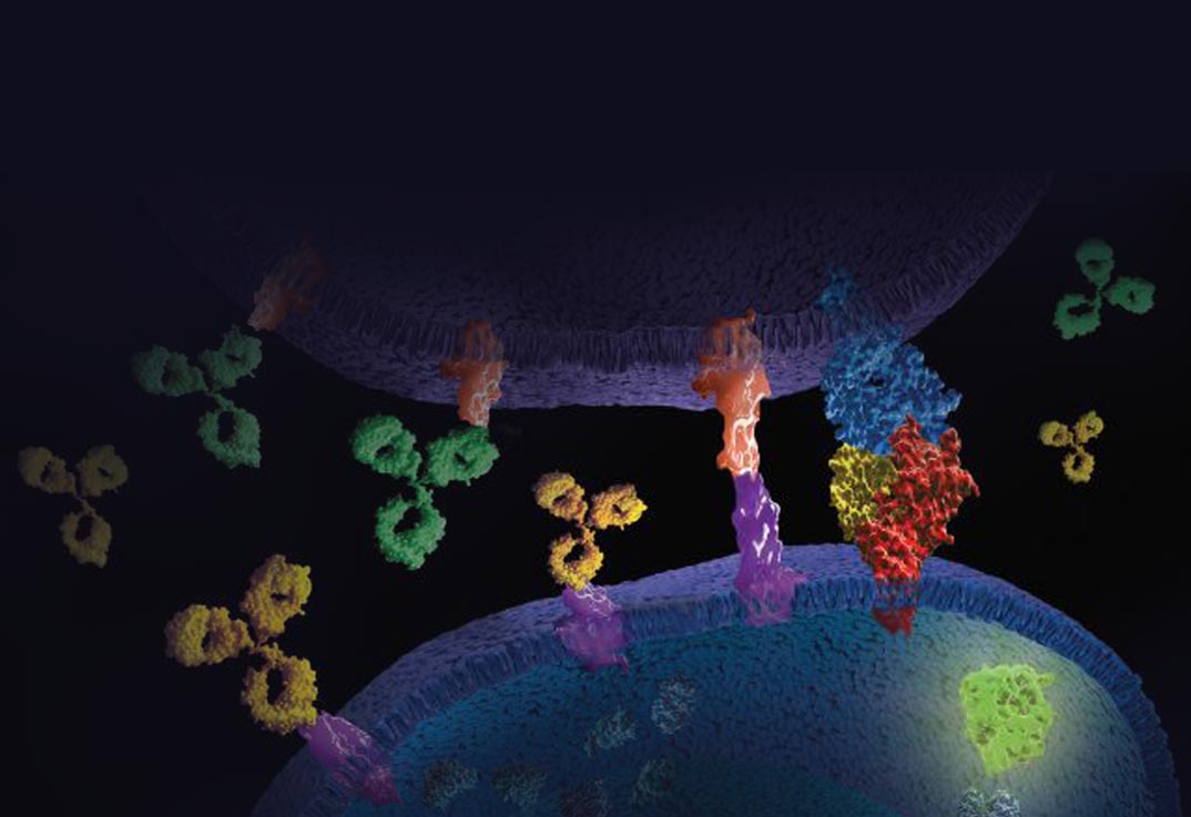
T cell activation is initiated by engagement of the T cell antigen receptor (TCR)/CD3 complex and the co-stimulatory receptor CD28. Engagement of TCR/CD3 on the cell surface leads to intracellular signaling events and the activation of nuclear transcription factors such as Nuclear Factor of Activated T cells (NFAT). In addition, co-engagement of TCR/CD3 with the co-stimulatory receptor CD28 leads to activation of AP-1 and NF-kB pathways. The IL-2 promoter contains DNA binding sites for NFAT, NF-kB and AP-1. Therefore, co-engagement of TCR/CD3 and CD28 results in IL-2 production, which is commonly used as a functional readout for T cell activation.
Traditional methods used to measure TCR-mediated T cell proliferation and cytokine production rely on laborious and highly variable primary peripheral blood mononuclear cells (PBMCs). However, functional reporter bioassays are now available that overcome limitations of PBMC-based assays by providing a workflow that is simple, reproducible, and scalable for antibody screening and drug discovery.


